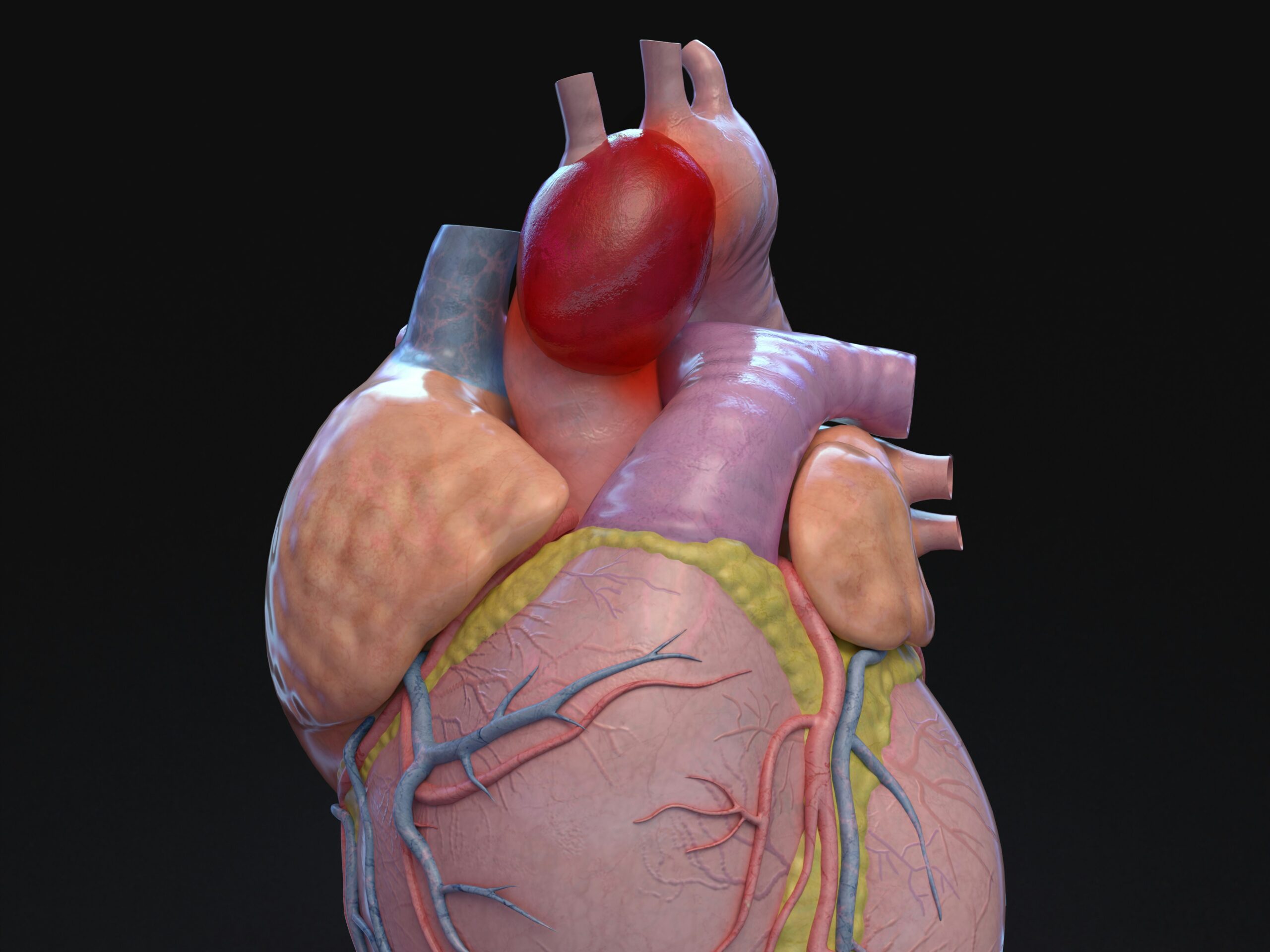
Emerging Imaging Techniques in Cardiology Explained

Modern cardiology faces increasing challenges as heart disease grows more complex and diverse. Traditional imaging methods like echocardiography and standard angiography remain valuable, but they sometimes fail to capture subtle or unusual abnormalities. Emerging imaging techniques in cardiology are transforming how specialists detect, evaluate, and manage complex heart conditions. These innovations provide detailed insights into cardiac structure, function, and blood flow, allowing clinicians to make precise, timely decisions.
By integrating advanced technology with clinical expertise, cardiologists can now visualize the heart in ways that were previously impossible. Three-dimensional imaging, high-resolution CT scans, magnetic resonance imaging (MRI), and nuclear cardiology techniques reveal details that enhance both diagnosis and treatment planning. Patients benefit from more accurate evaluations, less invasive procedures, and tailored therapeutic approaches that improve long-term outcomes.
The Role of Advanced Echocardiography
Echocardiography has long served as a cornerstone of cardiac imaging, but advanced techniques are expanding its diagnostic power. Speckle-tracking echocardiography, for instance, measures myocardial strain and detects early changes in heart muscle function that conventional echocardiograms might miss. This technique allows clinicians to identify subtle damage in patients with heart failure, cardiomyopathies, or chemotherapy-induced cardiac stress.
Three-dimensional echocardiography also provides a more complete view of cardiac chambers, valves, and blood flow patterns. By rotating and analyzing the heart from multiple angles, cardiologists can assess structural abnormalities with greater precision. This capability is especially useful in planning interventions such as valve repair, device implantation, or surgical correction. As a result, emerging imaging techniques in cardiology enhance both diagnostic accuracy and treatment effectiveness.
Cardiac Computed Tomography (CT) in Complex Cases
Cardiac CT has evolved beyond basic coronary artery evaluation to address more intricate diagnostic needs. With high-resolution imaging, CT scans can reveal plaque composition, coronary anomalies, and even myocardial perfusion patterns. These insights help physicians assess the risk of heart attack, identify vulnerable plaques, and guide preventive strategies.
Additionally, CT angiography enables detailed mapping of coronary arteries without the need for invasive catheterization in many patients. When combined with advanced software algorithms, these scans allow cardiologists to simulate blood flow, predict obstruction effects, and plan interventions with greater confidence. By incorporating CT into complex case evaluations, emerging imaging techniques in cardiology provide a non-invasive yet highly informative alternative for patients at risk.
The Impact of Cardiac Magnetic Resonance Imaging (MRI)
Cardiac MRI has become indispensable for evaluating complex structural and functional heart disorders. Unlike other imaging modalities, MRI offers high-contrast images of soft tissue, enabling precise assessment of myocardial scarring, fibrosis, and inflammation. This detail is particularly important for diagnosing cardiomyopathies, myocarditis, and congenital heart defects that may otherwise remain undetected.
Furthermore, MRI can quantify blood flow, chamber volumes, and ventricular function, providing a comprehensive picture of cardiac performance. When combined with contrast agents, it highlights areas of delayed enhancement, pinpointing regions of damaged tissue. These capabilities allow clinicians to develop targeted treatment plans and monitor disease progression over time. Consequently, MRI exemplifies the power of emerging imaging techniques in cardiology to transform complex case management.
Nuclear Imaging and Functional Assessment
Nuclear cardiology techniques, including positron emission tomography (PET) and single-photon emission computed tomography (SPECT), add another dimension to cardiac evaluation. These tools assess myocardial perfusion and metabolic activity, helping identify areas of ischemia or tissue viability that structural imaging alone may not reveal.
PET imaging, in particular, provides precise quantification of blood flow and detects subtle perfusion defects. This capability assists in evaluating patients with multi-vessel coronary disease, microvascular dysfunction, or unexplained angina. By integrating functional data with anatomical imaging, nuclear cardiology enhances clinical decision-making and guides interventions. Overall, these techniques exemplify the broader trend of emerging imaging techniques in cardiology, combining structure, function, and metabolism to achieve more comprehensive diagnoses.
Hybrid Imaging and Multimodal Integration
Hybrid imaging systems, such as PET/CT or PET/MRI, combine anatomical and functional data in a single session. These approaches allow physicians to correlate structural abnormalities with perfusion or metabolic changes, providing unparalleled insight into complex heart conditions. This integration reduces diagnostic uncertainty and enhances the precision of treatment strategies, particularly for patients with coronary artery disease, cardiomyopathies, or complex congenital anomalies.
Multimodal imaging also enables pre-procedural planning for interventions such as stenting, bypass surgery, or device implantation. By offering a detailed roadmap of the patient’s cardiac anatomy and function, hybrid techniques reduce procedural risk and improve outcomes. This trend underscores how emerging imaging techniques in cardiology increasingly rely on combining modalities to deliver comprehensive and actionable information.
Minimally Invasive and Real-Time Imaging
Real-time imaging has transformed both diagnosis and interventional cardiology. Techniques such as intracardiac echocardiography (ICE) and three-dimensional rotational angiography provide live, high-resolution views during procedures. These technologies guide catheter placement, device deployment, and structural repairs with remarkable precision.
By visualizing the heart in motion, cardiologists can adjust strategies immediately, reduce complications, and enhance procedural success. Real-time imaging also decreases the need for repeated interventions and shortens recovery times. These advancements exemplify how emerging imaging techniques in cardiology are bridging the gap between diagnostic evaluation and therapeutic execution, ensuring safer and more efficient care.
Benefits of Adopting Emerging Imaging Techniques
The adoption of advanced imaging methods offers several advantages. First, they improve diagnostic accuracy, allowing early detection of conditions that may otherwise remain hidden. Second, they facilitate personalized treatment planning by revealing the precise location and severity of abnormalities. Third, many techniques are non-invasive or minimally invasive, reducing patient discomfort and procedural risk.
Patients with complex cardiovascular conditions particularly benefit from these technologies. By combining multiple imaging modalities, clinicians can create detailed maps of the heart and vascular system, detect subtle dysfunctions, and monitor progression over time. The comprehensive data gained from these approaches support informed decisions, targeted interventions, and improved long-term outcomes.
Challenges and Considerations
Despite their advantages, emerging imaging techniques in cardiology present challenges. Cost, accessibility, and the need for specialized training can limit widespread use. Additionally, some imaging methods require contrast agents or radiation exposure, which may not be suitable for all patients. Physicians must weigh the benefits of advanced imaging against these factors while maintaining patient safety.
Ongoing research seeks to address these limitations by developing safer contrast agents, lower-dose imaging protocols, and more portable equipment. Education and collaboration across multidisciplinary teams further ensure that these technologies are used appropriately and effectively. As adoption expands, these challenges are gradually being mitigated, making advanced imaging a realistic option for a broader patient population.
Future Directions in Cardiac Imaging
The future of cardiology imaging promises even greater precision and efficiency. Artificial intelligence (AI) and machine learning algorithms are being integrated into imaging analysis to identify patterns and predict disease progression. AI can enhance image interpretation, reduce human error, and provide decision support for complex diagnoses.
Additionally, wearable and portable imaging devices are on the horizon, enabling continuous cardiac monitoring and remote evaluation. These innovations, combined with hybrid and multimodal imaging, will allow clinicians to deliver truly personalized care. As research advances, emerging imaging techniques in cardiology will continue to redefine the standard of care, improving early detection, treatment accuracy, and patient outcomes.