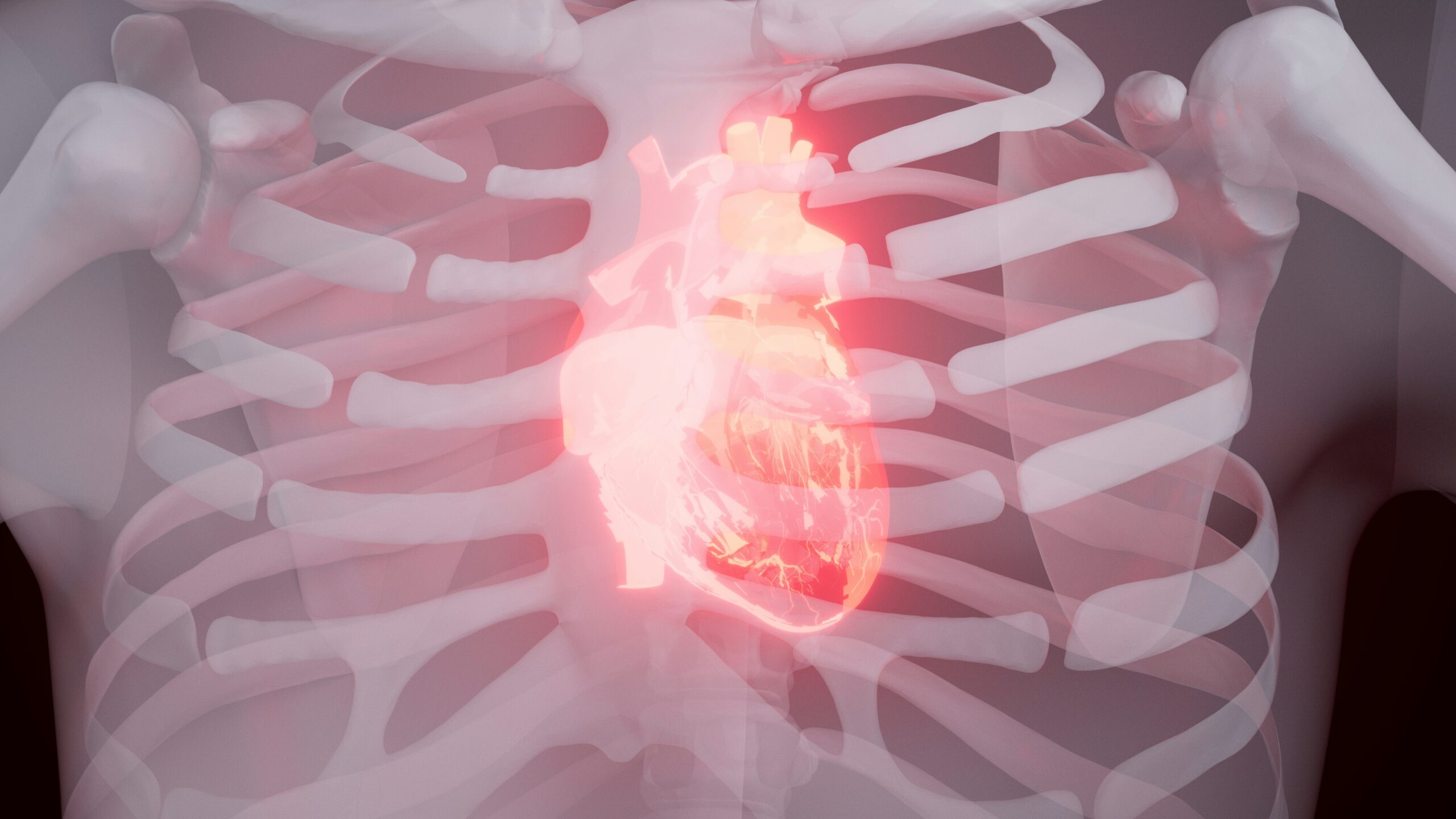
Beyond the Stethoscope: Emerging Imaging Techniques in Cardiology for Complex Diagnoses

Cardiology has always been at the forefront of medical innovation, but today’s advances in imaging are transforming how doctors uncover, understand, and treat heart disease. Gone are the days when the stethoscope and a basic X-ray told most of the story. Now, cutting-edge imaging tools are helping cardiologists see the heart with remarkable clarity—revealing details that were once invisible.
In this article, we’ll explore the most promising imaging techniques that are changing the landscape of cardiac care, especially in diagnosing complex cases where every detail matters.
Why Imaging Matters More Than Ever
Heart disease is often called the “silent killer” because its symptoms can be subtle until it’s too late. Imagine bridges that gap between uncertainty and clarity. The right scan can reveal blocked arteries, hidden structural problems, or subtle changes in heart tissue before they escalate into emergencies. For patients, that means earlier intervention, fewer invasive procedures, and more peace of mind.
3D Echocardiography: Bringing Depth to Diagnosis
Traditional echocardiograms provide two-dimensional pictures of the heart, but 3D echocardiography adds an entirely new perspective. By capturing real-time, three-dimensional images, doctors can evaluate heart valves and chambers with unmatched accuracy.
For example, when surgeons plan a valve repair, 3D echocardiography helps them visualize the exact shape and movement of the valve. It’s like upgrading from a paper map to Google Earth—it offers depth, detail, and context that can make or break a treatment plan.
Cardiac MRI: The Gold Standard for Tissue Clarity
When doctors need to understand the heart’s tissue in detail, cardiac MRI is often the go-to tool. Unlike CT scans, it doesn’t use radiation and provides crystal-clear images of both structure and function.
One of the most powerful uses of cardiac MRI is in diagnosing conditions like myocarditis (inflammation of the heart muscle) or differentiating between healthy and scarred tissue after a heart attack. Patients often describe the experience as lengthy—lying still inside a large scanner—but the payoff is a precise, detailed picture that helps avoid unnecessary procedures.
CT Angiography: A Non-Invasive Look at the Arteries
For decades, the only way to get a clear view of coronary arteries was through invasive catheterization. Now, CT angiography offers a safer alternative. With just an intravenous dye and a high-speed CT scan, cardiologists can map the arteries in astonishing detail.
This technique is particularly valuable for patients with chest pain where the cause isn’t immediately clear. Instead of rushing to the catheter lab, doctors can use CT angiography to rule out major blockages, sparing patients from unnecessary procedures.
Nuclear Imaging: Tracing Blood Flow with Precision
Sometimes, the key question isn’t what the heart looks like, but how well it works. Nuclear imaging techniques, like PET and SPECT scans, use small amounts of radioactive tracers to follow blood flow through the heart muscle.
These tests are especially useful for patients who may have coronary artery disease but have normal results on other imaging. By revealing “silent” areas of reduced blood flow, nuclear imaging helps ensure no problem is overlooked.
Fusion Imaging: When One Picture Isn’t Enough
The future of cardiology isn’t about replacing one imaging method with another—it’s about combining them. Fusion imaging merges data from different scans, like echocardiography and CT, into a single, unified view.
Imagine a surgeon preparing for a complex heart procedure. Instead of flipping between images from multiple machines, they can see a layered, comprehensive picture that blends structural and functional details. It’s like overlaying street maps with traffic data before heading out on a drive—smarter, safer, and more efficient.
Artificial Intelligence: The Silent Assistant Behind the Scenes
AI is making waves across medicine, and cardiology is no exception. Advanced algorithms can now analyze imaging scans in seconds, flagging subtle abnormalities that even experienced eyes might miss.
For patients, this doesn’t mean robots are replacing doctors. Instead, AI acts as a second set of eyes, boosting accuracy and reducing diagnostic errors. In busy hospitals, it also speeds up the process, meaning quicker results and faster treatment decisions.
What It Means for Patients and the Future
For patients, these emerging imaging tools translate into more personalized, less invasive care. A person with unexplained chest pain might avoid an unnecessary surgery thanks to CT angiography. Someone recovering from a heart attack may get a clearer recovery plan through cardiac MRI. And a patient at risk for sudden heart failure could receive life-saving insights through nuclear imaging.
Looking ahead, as these technologies become more affordable and accessible, even community hospitals will be equipped to handle complex diagnoses with confidence. The ultimate goal isn’t just better pictures of the heart—it’s better outcomes for the people who rely on it every day.
Final Thoughts
Emerging imaging techniques in cardiology aren’t just technological marvels—they’re tools of compassion. By allowing doctors to see more, earlier, and more clearly, these advances are rewriting the story of heart disease.
The future of cardiac care will likely blend human expertise with cutting-edge imaging and AI, giving patients not only longer lives but healthier, more hopeful ones. And that’s a vision worth keeping close to the heart.
Additional Information
- Blogs
- Nishi Patel